An unstable ankle rarely happens overnight. It develops from a rolled ankle that never quite healed, a season of playing through pain, or years of uneven ground and worn ligaments. By the time someone walks into a clinic with recurrent sprains, swelling after routine activity, and that constant sense of “giving way,” the mechanics of the ankle have changed. The lateral ligaments, especially the anterior talofibular ligament and calcaneofibular ligament, are stretched or torn. The body compensates with stiff calf muscles, guarded gait, and subtle shifts in foot posture that spread stress elsewhere. A foot and ankle surgeon looks beyond the single sprain, mapping the chain of movement from hip to toes to understand why the ankle keeps failing and how to fix it. Two of the most reliable solutions, the Broström repair and an internal brace augmentation, anchor a modern approach to stability that preserves normal motion and shortens recovery when chosen well.
What a foot and ankle specialist actually evaluates
An effective consult goes far past “does it hurt here.” A foot and ankle orthopedic surgeon or foot and ankle podiatric surgeon will check laxity with ankle inversion stress and anterior drawer tests, compare side to side, and feel for tenderness along the fibula, sinus tarsi, and peroneal tendons. We watch how you walk, not just on a straight hallway but with turns, on your toes and heels, and sometimes on a treadmill to see fatigue patterns. Strength testing focuses on the peroneal muscles, tibialis posterior, and intrinsic foot stabilizers. Range of motion is measured in degrees, noting capsular stiffness versus muscular tightness. If the ankle clunks with a certain motion or there is a crunching sensation under the lateral malleolus, we consider peroneal tendon subluxation or osteochondral defects of the talus.
Imaging fills in the missing puzzle pieces. Weightbearing radiographs show alignment and any bony contributions to instability. Stress radiographs can quantify laxity, although the physical exam is often enough in experienced hands. If symptoms persist or mechanical clicking suggests cartilage injury, an MRI helps identify partial versus complete ligament tears, peroneal tendon split tears, scarring in the sinus tarsi, and bone marrow edema. A CT scan may be useful before revision surgery or when we suspect subtle malunions from prior fractures.
Patterns matter. A varsity soccer player with a high-velocity inversion injury and immediate swelling is different from a hiker with ten minor sprains over three years. The first often needs timely intervention to protect cartilage. The second may improve with focused rehabilitation but risks progressive laxity if the ligaments have lengthened too far. The art is sorting who responds to physical therapy and bracing and who benefits from surgical repair.
Conservative care first, when it works
Not every unstable ankle needs an operating room. In many patients, a structured plan corrects muscle timing and restores neuromuscular control. I typically start with a six to eight week program that includes balance work on unstable surfaces, peroneal strengthening both concentrically and eccentrically, and calf and hamstring flexibility. Foot posture gets just as much attention as ankle strength. Flatfoot posture shifts the talus inward and challenges the lateral ligaments, while a high-arched rigid foot can increase inversion moments. A foot and ankle biomechanics specialist may suggest a semi-rigid orthotic for valgus rearfoot control or a lateral wedge for a cavus foot to soften the inversion lever.
Bracing has a place, especially during return to sport. A lace-up ankle brace or figure-8 style strap reduces recurrent sprains in at-risk athletes. Taping can help in the short term, though skin tolerance and time make it a poor long-term strategy. Anti-inflammatories, topical or oral, settle the synovitis that often lingers around an unstable joint. If the ankle continues to give way despite faithful therapy, or if imaging shows poor tissue quality, the conversation shifts to repair.
The Broström repair in plain terms
The Broström is the workhorse procedure for chronic lateral ankle instability. It is a direct repair of the native ligaments back to the fibula, often with a Gould modification that advances https://www.google.com/maps/d/u/0/embed?mid=1KK5h61ew-Y56FlIWVfZTUt-LKxfgWdo&ehbc=2E312F&noprof=1 the extensor retinaculum for added support. The beauty of this approach is that it restores anatomy rather than replacing it with a tendon graft. When tissue quality is adequate, it provides stability with normal mechanics.
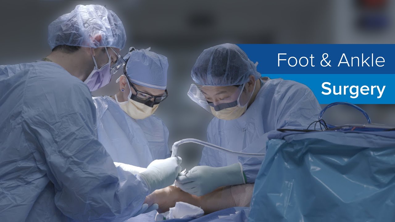
In the operating room, the incision sits just anterior to the fibula. The torn ends of the anterior talofibular ligament and calcaneofibular ligament are identified, cleaned up, and anchored back to the fibula with suture anchors. The retinaculum is then advanced over the repair, reinforcing the construct. If the peroneal tendons look inflamed or frayed, they can be debrided or repaired at the same sitting. Cartilage lesions discovered on imaging or arthroscopic inspection can also be addressed, from microfracture for small defects to grafting in select cases.
Most foot and ankle surgery experts perform the Broström through a small open incision, though some steps can be done arthroscopically. Open repair allows direct handling of tissue, which remains the most predictable method in my hands, particularly when there is scarring or prior surgery.
Where the internal brace fits
An internal brace is not a new ligament. It is a small, braided suture tape that spans from the fibula to the talus and often to the calcaneus, fixed with low-profile anchors. Think of it as a seatbelt that protects a newly repaired ligament while it heals and helps resist early inversion forces. It is invisible from the outside and adds minimal hardware.
When do I add it? Three situations come up repeatedly. First, Rahway, NJ foot and ankle surgeon high-demand athletes who plan to pivot, cut, and jump within months of surgery. Second, patients with generalized ligamentous laxity or poor tissue quality, where a standard Broström alone may stretch out. Third, revision cases after a failed prior repair. The internal brace reduces early elongation of the repair and gives confidence in the first weeks of motion. In biomechanical testing, constructs with a suture tape augmentation resist more inversion torque before failure compared to suture repair alone. In clinic, that translates to fewer early re-sprains and a quicker progression of activity, though final outcomes in everyday patients are similar at one to two years.
Internal bracing does not replace sound judgment. Over-tightening can limit normal ankle motion, especially plantarflexion-inversion, and can cause discomfort with certain movements. Placement must respect the ligament footprints. The anchors should be low profile and oriented to align with native fiber direction. When executed well, patients often regain controlled motion faster, while the repair maintains stability.
What recovery looks like, week by week
Patients ask for exact timelines. Every foot and ankle treatment doctor knows biology does not read calendars, but a thoughtful pathway helps set expectations. Tissue quality, age, cartilage health, and job demands shape the pace.
- An example pathway after a Broström with internal brace: Week 0 to 2: Splint or short leg cast. Foot elevated above heart level as much as possible. Non-weightbearing or touch-down only for balance in the bathroom. Focus on swelling control and gentle toe curls to prevent stiffness. Week 2 to 6: Move into a walker boot. Begin partial weightbearing and progress to full as comfort allows, usually by week 3 or 4. Start gentle dorsiflexion and plantarflexion within a protected range. Stationary bike with the boot on is allowed in many cases. Sutures come out around day 10 to 14. Week 6 to 10: Transition from boot to a supportive shoe with an ankle brace. Begin supervised balance work, resisted peroneal strengthening, and closed chain exercises. Light jogging on level ground may begin around week 8 to 10 in selected patients. Week 10 to 16: Agility progressions, cutting and pivoting drills, sport-specific work. Most recreational athletes return between 12 and 16 weeks. Elite athletes may return earlier with careful testing, though I prefer strength and balance symmetry within 10 percent before full clearance.
Without an internal brace, the early phases are more conservative, and running often begins closer to week 12. Desk workers usually return around two weeks in a boot. Physically demanding jobs that involve ladders, uneven ground, or carrying heavy loads often require 8 to 12 weeks, sometimes more.
The patient stories that teach us
A distance runner came to clinic after three years of repeated ankle rolls, always around mile seven on trails. Exam showed grade 2 laxity, and MRI revealed an attenuated anterior talofibular ligament with a small osteochondral lesion of the talus. We performed a Broström with internal brace and microfracture of the lesion. He respected the early boot phase, did his balance work religiously, and we held off trail running until he could hop on one leg for 60 seconds without wobble. He finished a half-marathon at six months with no ankle fear. The internal brace likely helped him tolerate early strengthening without stretching the repair.
A collegiate volleyball player sprained her ankle just before playoffs. Exam was consistent with a high-grade tear, and MRI showed poor tissue quality and a split peroneus brevis tendon. After discussing options, we repaired the ligament with internal brace augmentation and addressed the tendon. She returned to practice drills at 10 weeks and played limited minutes at 12. The tendon repair mattered as much as the ligament stabilization, a reminder that the lateral ankle is a neighborhood, not a single house.
Choosing Broström, internal brace, or both
The decision matrix includes patient goals, tissue quality, ligament damage pattern, and history of prior surgery. Primary instability with good tissue often does well with a Broström alone, but if an athlete wants to shorten the guarded period or has ligamentous laxity, the internal brace is a smart adjunct. Revision surgery or cases with generalized hypermobility benefit from the extra checkrein. In rare cases with severe attenuation or absent tissue, we discuss tendon graft reconstruction, usually with a peroneus longus split or hamstring autograft or allograft. That option sacrifices a bit of normal kinematics to gain stability and is reserved for complex cases.
A foot and ankle ligament specialist should also scan for coexisting problems. A cavus foot with a tight lateral column predisposes to recurrent inversion. In that context, a soft tissue release or small osteotomy may be necessary to protect the repair. Peroneal tendon instability can masquerade as ligament laxity and may require retinacular repair. Persistent deep ankle pain after a sprain hints at an osteochondral lesion, which we should address during the same anesthesia to avoid future surgeries.
Risks and trade-offs, spoken plainly
Surgery is predictable, not perfect. Infection rates are low, usually well under 2 percent with standard precautions. Nerve irritation, particularly of the superficial peroneal nerve, can cause patches of numbness or sting along the incision. Most cases settle over weeks to months. Stiffness is uncommon with modern rehab, but overly aggressive early motion or premature return to cutting sports can stretch the repair. Hardware irritation from anchors is rare with current implants, but occasional sensitivity occurs in very thin patients.
The biggest risk is underestimating the underlying mechanics. Repairing a ligament without addressing foot posture or tendon pathology may buy a year of stability rather than a decade. Conversely, over-tightening a repair can protect against inversion but restrict smooth ankle motion, which athletes notice during landing and quick pivots. Judicious tensioning while the foot is positioned neutrally minimizes this.
What your first appointment should cover
The most productive visits are collaborative. Come prepared to describe the number of sprains, any sense of locking or catching, and what activities provoke symptoms. Bring prior imaging. A foot and ankle pain doctor will examine both ankles, check alignment, and discuss the spectrum from therapy to surgery. If you are a candidate for repair, insist on a clear recovery plan, including brace use, therapy milestones, driving limitations, and return-to-work timing tailored to your job.
Here is a concise checklist you can use to gauge the completeness of the plan:
- Do we agree on the diagnosis and any coexisting issues like peroneal tendon tears or cartilage injury? Why is a Broström, an internal brace, or both recommended in my case? What is the week-by-week plan for weightbearing, range of motion, and therapy? How will my sport, job, or foot posture influence the approach and recovery? What are the contingency plans if healing is slower or symptoms persist?
Rehabilitation that respects biology
Strong repairs deserve smart rehab. Early on, the priority is quieting the ankle: swelling control, gentle motion within a protected arc, and safe progression of weightbearing. As healing progresses, we emphasize three components that separate durable outcomes from fleeting ones.
First, proprioception, the body’s ability to sense position, must be retrained. Wobble boards, single-leg stands with eyes closed, and dynamic balance tasks on foam or BOSU surfaces matter more than calf raises. Second, eccentric strength of the peroneals protects against sudden inversion. Controlled lowering drills with resistance bands and step-down tasks build resilience. Third, movement quality during cutting needs deliberate practice. Ladder work, cone drills, and sport-specific landings, supervised initially, smooth out the stiff, protective patterns that patients adopt after months of instability.
A foot and ankle motion specialist or experienced physical therapist can tailor this work. Good therapists also watch for compensation higher up the chain. Hip abductors and external rotators often weaken with prolonged ankle problems, shifting loads to the ankle and knee. Reinforcing hip strength and trunk control stabilizes the entire limb.
How surgeons individualize technique
Two surgeons might both perform a Broström with internal brace and yet make different intraoperative choices. That reflects judgment, not inconsistency. Anchor selection may vary based on bone quality. Suture tape placement is adjusted when the talar footprint is small or when the calcaneofibular ligament needs more emphasis for patients who struggle on uneven ground. If the peroneal tendons are tight within a narrow groove, a foot and ankle tendon specialist may open and deepen the groove to prevent snapping. Each decision adds a few minutes but can prevent years of annoyance.
For patients with generalized laxity, the retinacular reinforcement is more than a courtesy layer. It provides a broader sheet of collagen to resist inversion. In revision cases, scar tissue can blur anatomy. Here, an experienced foot and ankle surgical expert takes time to identify healthy edges and may add an allograft reinforcement if tissue quality disappoints, even when an internal brace is used.
Expectations for long-term outcome
Most patients regain stable, confident ankles. Studies and daily experience align: when performed for the right indications, more than 85 to 90 percent of patients report improved stability and function. Recreational athletes return to running and field sports. Hikers reclaim uneven trails. Occasional weather-related aches happen after long days or in cold conditions, but they are a nuisance rather than a limitation. The repair protects cartilage by reducing abnormal shear, though prior injuries may still produce intermittent stiffness.
Footwear choices and terrain still matter. A minimalist shoe on rocky trails invites trouble, even with a well-healed repair. For those who love aggressive sports, a supportive shoe with a stable heel counter and, during the first year, an ankle brace for games remains a wise habit. Most of my high school and college athletes keep a brace in their gym bag long after discharge from care, using it during tournaments or when fatigue sets in.
How this compares with other options
Tendon graft reconstructions, such as a Chrisman-Snook or an anatomic ligament reconstruction with hamstring autograft, still have a place. They are reserved for severe laxity, failed repairs with absent tissue, or when additional stability is required for heavy laborers with high-risk environments. They trade slightly less normal kinematics for stronger static restraint. Recovery often runs longer, and there is a small risk of donor site symptoms if an autograft is used.
Arthroscopic thermal capsular shrinkage had a moment years ago but fell out of favor due to high failure rates. Platelet-rich plasma injections have limited value for chronic, elongated ligaments, though they may help with associated tendinopathy. Bracing alone can be reasonable for low-demand patients or those with medical risks that make surgery unwise, but repeated sprains still threaten cartilage and peroneal tendons over time.
The team around your ankle
A good outcome is a team sport. The foot and ankle healthcare provider leads, but success leans on a skilled physical therapist, a shoe specialist who understands foot shape and activity demands, and sometimes a coach willing to modify drills during return. Communication matters. If your job involves ladders, we plan around that. If you manage diabetes, a foot and ankle diabetic foot specialist coordinates wound care and glycemic control to protect healing. If nerve pain complicates recovery, a foot and ankle nerve specialist can help with targeted therapies.
A foot and ankle medical expert brings more than surgical skill. We are pattern recognizers and problem solvers. The Broström and internal brace are strong tools, but the right plan wraps them in careful diagnosis, thoughtful rehab, and realistic expectations. When those elements align, ankles that once felt unreliable become boring again, which in this context is the best compliment of all.
When to seek a dedicated foot and ankle surgeon
If you are on your third sprain in a year, if your ankle clicks and swells after routine errands, or if you avoid stairs because the joint feels loose, it is time to see a foot and ankle specialist. Look for a foot and ankle orthopedic surgeon or foot and ankle podiatric physician with a steady volume of ligament repairs and sports injuries, someone who can articulate the differences between a Broström alone and an internal brace augmentation and who examines the whole limb, not just the painful spot. Ask about outcomes, revision rates, and how they tailor rehab to your sport or work.
The best first step is simple: an honest evaluation, clear options, and a recovery plan you believe in. From there, whether your path runs through a strong round of therapy or a precise Broström with an internal brace, the goal is the same. Stability, confidence, and the freedom to move without thinking about your ankle.